Monday, July 26, 2010
Week 5
Monday morning I came into the lab and began developing last weeks membranes for western analysis. I used DAX1 as my primary antibody, since the gel contained a lane of isolated DAX1. Unfortunately, the IEF gel markers were the only bands to appear. At this point, neither Dr. Clendening or I had any idea as to why the transfers were unsuccessful.
Phone Call
Dr. Clendening and I decided it would be best to call up the company that provided us with our IEF gel and try to troubleshoot. The woman that I spoke with recommended using the acetic acid transfer, but letting it go overnight. She said that she would send us a gel right away.
IEF Again
When the IEF gel came in, we decided to run last week's experiment again. Samples of the nuclear fraction, nuclear fraction with formaldehyde/glycine treatment, and isolated DAX1 were loaded into the gel. Experimental conditions were kept constant, except we ran the transfers overnight. I developed the westerns the following day, but we got the same result as before. DAX1 was not detected in any of the samples.
Friday, July 23, 2010
*Greenhouse Greatness!*
I also began the revisions to the methods of the bulbil dispersal experiment to make sure that there was no room for error. I created all new data sheets, used all new bulbils and set up stations to ensure that everything would run smoothly. I had two assistants that helped me and we began the project last week. This week, I completed the bulbil dispersal experiment and began to enter all of the new data. I will begin to analyze all of the data next week with the help of Dr. Sanford and Dr. Aronson.
On Tuesday, Dr. Aronson and I went to Alley Pond in New York City to help with a Forest Restoration project . Although, I am always looking forward to being out in the field, we were all eaten alive by malicious mosquitoes, literally! On Thursday, we went to a different site at Roy Wilkins Park in Queens to assist on the same project. This site was way more pleasant and we got a lot accomplished. We have some upcoming sites that I am looking forward to. This is such a great partnership! On Friday, Dr. Aronson and I went over to the high school to check on my plants. Everything looked pretty good and no plants have germinated as yet, except for two weeds. I will return next week to check on them again.
Wednesday, July 21, 2010
Cardiology Fellowship... EP lab and Consult SVC
In the beginning of each week the Cardiology team meets for a morning conference to discuss how the weekend went, and makes plans for the week to come. On the first day of my internship I started out meeting everyone at the morning conference then jumped right into a bunny suit, which is a sterilized outfit that allows me to observe procedures without possibly harming the patient, and watched my first generator change in the EP lab. It really fascinated me to see how small such an important device can be and that the procedure is done quite quickly. Throughout the rest of the week I observed a few pacemaker implantations and a cardio version as well. For those of you that don’t know what a cardio version is, it occurs when a patient has an irregular heart rate, and this procedure is done to shock the heart in an attempt to reset their heart rate to a safe rhythm. I also followed a Nurse Practitioner to observe the post procedure and learned how defibrillators and pacemakers are controlled.
The next service I followed was the Cardiology Consult service. In the cardiology consult service I learned about the many reasons people are placed under Cardiologists care, and the different tests done in order to determine the best source of treatment for the patient. One interesting procedure I learned about was hypothermia treatment for patients that have a cardiac episode; the hypothermia treatment is performed by cooling the body after stabilizing the patient’s heart in an attempt to preserve brain function, and has been shown to be quite effective. Throughout this week I also was able to listen to a patient’s heart that had a heart murmur, and feel their unique pulse.
These first couple weeks I have had a great time and have learned so much. I can’t wait to experience the next 8 weeks of the internship!
Jennalee Trombley
Sunday, July 18, 2010
Week 4
The beginning of the week was devoted to running an actual version of my experiment. I incubated samples of DAX1 and SF1 at male producing temperature (MPT), 26 degrees celsius, and female producing temperature (FPT), 31 degrees celsius. Half of the samples received a formaldehyde/glycine treatment to theoretically "fix" the proteins and all of the samples were loaded into a 4-20% polyacrylamide gel. The gel ran for an hour at 100 volts. Afterwards, the proteins were transferred to nitrocellulose membranes through electroblotting (I electroblotted twice to get two, presumably, identical membranes).
The next day I developed western blots, but the results were not satisfactory. Half of the samples did not show up (left side of photos). The lanes that did appear reacted with both DAX1 and SF1 antibodies, but these lanes should have reacted with only one or the other. It seemed as though there might have been some non-specific binding of the antibodies, so Dr. Clendening suggested that I lower the concentration of antibody in the future. We had no explanation for the lanes that did not show up. Perhaps the proteins got degraded?
I spent the middle of the week researching the protein Wilm's Tumor Suppressor (WT1). I had run out of gels, so I had to find something to do in the meantime. It turns out that WT1 plays a role in TSD. It has many different isoforms, which may or may not be regulated by temperature. WT1 would serve as a prime candidate for future experimental applications.
IEF Test Run
By the end of the week the IEF gels had arrived and I was able to return to the DAX1/SF1 experiment. I ran an IEF gel on Friday with samples of nuclear fraction, nuclear fraction with formaldehyde/glycine treatment, and DAX1 isolated. I tried three different methods for transferring: electroblot with acetic acid buffer, electroblot with tris-glycine buffer, and a pressure blot with tris-HCl buffer. The membranes were left to dry over the weekend.
Saturday, July 17, 2010
June 16
Later that day when the sun started to go down a bit we went back out to try and pick out exactly where we want our plot. We decided on a fairly dense patch of wood that was barely disrupted by Forestry. Tomorrow we will get the coordinates and hopefully get started.
Friday, July 16, 2010
*Revelations in my Research!*
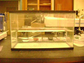
Ultimately, I calculated the volume, density and buoyancy of these bulbils. I met with Dr. Sanford to discuss what was next in terms of the project and I got started on it right away. I am testing whether my prediction of buoyancy is an accurate measure of the metric in water. I want to simulate how bulbils will sink in a stream, so I used a 10 gallon tank and filled it half way with water. I made sure that there were no currents in the water and got a precise measure of the depth of water. I was very careful to avoid the meniscus effect. I had an assistant with me to calculate the time it took to sink from the surface of water to the bottom of the tank. I was very careful not to touch the water too much by using a long pair of forceps so that the level of water remained the same.
I predicted that the bulbils would have a negative buoyancy which means that they would sink. Most of them did but a lot of them did not. I left them over night to see if anything would happen but unfortunately, I returned to the same results the following morning. The most logical reasoning for the bulbils not sinking was the fact that they began to lose water in the zip lock bags that they were enclosed in, in the freezer. I noticed that when I took the weights of the bulbils after I retrieved them from the water, they actually lost about half of their original mass. Therefore, Dr. Aronson and I decided that we would start the whole experiment over with fresh bulbils that we had stored. This time we agreed that we would do all of the measurements and timing of sinking at one time to prevent the bulbils from drying out between measurements. This is also all a part of the research process. Sometimes it means perfecting an experiment as many times as you need to, in order to ensure that it is successful. I am grateful for all of these experiences because it is helping me to become a better research student.
Myself along with Dr. Aronson, Mr. Weiss, and our Locust Valley High school student all went out to Prospect Park last week Friday to add another site to my diversity sampling of invaded and non-invaded areas. However, there was not much to sample from at this site due to the small size of the invasion and environmental conditions (Norway Maples casting deep shade).
Monday, July 12, 2010
*Getting Down at Muttontown!*
Jewel weed
After the sampling was completed, we collected soil from the non-invaded area as well as the invaded area. The soil will be used in the greenhouse experiment along with our activated carbon to test for allelopathic chemicals being left behind by this invasive species. I also decided on exactly which company I would order the activated carbon from by using scientific literature, searching on the web and contacting companies. We should have it in time for the greenhouse experiment.
We could not identify some of the species in the plots so we carried back samples from the field site. We could only identify one out of the six species that we were not sure about. We identified it by looking at identification books. This was the only species with flowers. However, we pressed the other species in the lab to save for later identification and we plan to return to the field site once these unknowns are flowering. It is very difficult to identify herbaceous plants that have not flowered yet.
Sunday, July 11, 2010
LIJ Cardiology Internship: Week 1 & 2
Every few days there is a morning conference at LIJ, and every Wednesday there is a lunch conference. They discuss things like interesting cases from over the weekend and unique EKG's that might be on the board exams. Sometimes presentations are given about the histories of certain procedures and the risk/benefit of one's that exist. The main thing I have observed is that there is no step by step guideline for the doctors to follow. Every patients situation brings it's own complications and the cardiologist has to factor everything together like age, allergies, chronic disease, family history, previous visits, and so on for each decision they make. It might seem like an implantable cardioverter defibrillator (ICD) should be introduced to someone because of frequent fribrillations, but they have a history of dementia and live alone in a trailer park. Some doctors might not see this as a ethical thing to do because the patient might not understand what is being put in them. There are so many decisions the cardiologists have to make in such intense situations.
My first week I spent on the cardiology consult service with one of the first year (now second year) fellows. This is a service where a few of the patients are directly under your care (primaries) and the rest are under the direct care of another physician and you are examining them only in terms of cardiology (consults). It is a very busy service, with a lot of patients, but each patient gets the time they need to be examined. After my week on the service, I was able to see the routine the fellow undergoes everyday which is to meet with the resident and get the vitals of each patient, and then go through each patients case with the attending physician to make a plan for the day. Once that was completed, we were off to visit each patient. Sometimes you would go to see a patient and they might be getting an Echocariogram or be in the Catheterization lab and you would have to come back. Sometimes they would be asleep and you would have to wake them up. At other times we would be going in to recommend procedures like valve replacement surgeries and pacemakers. Each patient has a story and a reason why they are there, and that needs to be understood before entering the room.
Dr. Slotwiner who organized this internship with Dr. Shanies, has been nothing but nice, helpful and motivating. Spending the week with him in the EP lab (electro-physiology) was a great experience. I got to stand in on an EP study and the implantation of a pacemaker device, which was one of the most amazing things I have ever seen. It is unbelievable how something so small can do so much! Not only can it pace the atrium and the ventricles separately, it understands threshold limits, and has a memory for when the heart has fribrillations and palpitations. I also was able to see an ablation, where an ectopic beat is corrected non-invasively!
My experiences thus far in the LIJ cardiology department have been nothing but excellent. I look forward to the weeks to come and to the new things I will learn.
-Stephanie Lombardi
Thursday, July 8, 2010
Week 3
The beginning of the week was focused on developing western blots from last week's wicking transfer. I followed the standard procedure for developing westerns, but only the IEF markers appeared. Furthermore, the markers were found on only one of the membranes, instead of both as expected. Dr. Clendening and I thought that the proteins may have been retained in the agarose gel, so we preformed a coomassie stain. Sure enough, there were proteins in the gel. We decided not to use agarose gels in the future.
In the middle of the week I tested the effects of the formaldehyde application. Last week's results indicated that the formaldehyde might have reacted with the extraction buffer from the NucBuster kit. To test this hypothesis, I isolated a nuclear fraction using a different protein extraction kit (presumably with different extraction buffers). This fresh sample was to be compared with a nuclear fraction from the previous week (NucBuster extraction kit). I aliquoted samples of the two nuclear fractions and gave them either the standard formaldehyde and glycine treatment or nothing at all. All of the samples were incubated on ice for 15 minutes. Upon examination, it was clear that the nuclear fraction from the NucBuster kit reacted with the formaldehyde/glycine application and formed a precipitate. The fresh sample, that was isolated from the other kit, did not appear to react. Dr. Clendening and I decided to continue with our test the next day and load samples from both extraction kits on a protein gel.
Yet, when I came into lab, there was a slight change of plans. Dr. Clendening wanted me to isolate nuclear fractions from fresh tissue using the NucBuster extraction kit. She also wanted me to isolate nuclear fractions from not only brain tissue, but also the adrenal/kidney tissue; the gel would hopefully tell us if one tissue was preferable over the other. I preformed the isolation steps and added the formaldehyde solution to half of the samples. But, when I was about to add the glycine, I noticed that it contained mold. Dr. Clendening thought that the mold might have caused the precipitate we had seen earlier in the week, so we made a new glycine solution and added it to the formaldehyde samples; it was unclear whether or not these samples formed any precipitate. We proceeded to load the samples into a 20-40% polyacrylamide gel and I successfully transferred the proteins to a nitrocellulose membrane later that day.
On Friday, I developed the western blot. Every sample showed up! Thankfully, we were able to learn many things from this gel. For one, the adrenal/kidney tissue contained more SF1 than the brain tissue. Secondly, the samples with formaldehyde/glycine were less visible than the untreated samples. They did show up though, so presumably the cross-linking experiment was feasible. Third of all, the isolated SF1 and DAX1 samples appeared, indicating that we had useful positive controls. Moreover, these samples could be used in later application to more accurately depict the interactions between SF1 and DAX1. Though, they would need to be concentrated, because they produced a faint band. Last of all, and perhaps most importantly, we recognized several bands of SF1 in the nuclear fraction. These bands indicated that SF1 was bound to some additional protein(s) or other nuclear component(s). Since our research was based upon the hypothetical binding of SF1 and DAX1, two different proteins, this evidence was rather encouraging.
Tuesday, July 6, 2010
Weeks 1 and 2
Most of week 1 was spent dissecting turtle embryos. The eggs, for the most part, had reached the temperature-sensitive period (TSP). Embryos were removed from the eggs and the brain, gonadal, and adrenal/kidney tissues were collected. These tissues were stored in a sodium/phosphate buffer and placed in the freezer at -80 degrees celsius.
Week 2
Week 2 was comprised of a variety of activities, all focused on determining the interactions between DAX1 and SF1. Like the previous week, a great deal of time was spent on collecting the necessary tissues. Eggs were dissected and the brain, gonadal, and kidney/adrenal tissues were stored in sodium phosphate buffer at -80 celsius. In addition, a nuclear fraction was extracted from a stored sample of brain tissue. This fraction was split into multiple aliquots, which were to be incubated and used in an isoelectric focusing (IEF) gel (since the fraction presumably contained both DAX1 and SF1).
Samples were incubated at 26, 31, and 0 degrees celsius. After 30 minutes, a formaldehyde solution was added to fix any protein-protein interactions that had taken place. Incubation continued for an additional 15 minutes, before a solution of 1.25 M glycine, pH 7.5, was added to stop the fixation process.
The IEF gel was loaded with the samples and run according to the gel electrophoresis recommendations. Proteins were transferred to a nitrocellulose membrane using acetic acid transfer and developed using the western blotting technique. Unfortunately, the results were not satisfying.
Only the IEF markers and the positive control (isolated SF1) appeared on the developed membrane. We were not sure why the others samples did not appear, but we did have a couple of predictions. First of all, we had never preformed an acetic acid transfer. The procedure was new to us and there were a number of things that could have gone wrong. Secondly, none of the samples treated with formaldehyde appeared. We had seen a white slurry form on top of the formaldehyde containing samples before loading, which we believed may have interfered. Our initial conclusion was that a salt had formed between the sodium phosphate buffer in the formaldehyde and the elution buffer in the nuclear extract.
To resolve our conflict we decided to test the transfer technique and make a new solution of formaldehyde. An agarose gel was made up this time and loaded with samples following the same conditions as before. The only difference was that the formaldehyde was made up in a tris-HCl buffer instead of sodium phosphate. The gel was run and transferred to nitrocellulose again, but the transfer conditions were altered. This time we decided to use the wicking method. We cut the gel in half, placed one half in acetic acid buffer, and placed the other half in saline-sodium citrate buffer (SSC).